

|
Drawings |
|
Syn.:
|
Anguillula dipsaci Kühn, 1857
Anguillula dipsaci (Kühn) Gerv. et. v. Ben., 1859 Tylenchus dipsaci (Kühn) Bastian, 1865 Anguillula devastatrix Kühn, 1868 Anguillula secale Nitschke, 1868 Anguillula putrefaciens Kühn, 1877 Tylenchus havensteini Kühn, 1881 Tylenchus hyacinthi Prillieux, 1881 Tylenchus alii Beijerinck, 1883 Tylenchus devastatrix Ritzema Bos, 1888 Ditylenchus phloxidis Kirianova, 1951 Ditylenchus fragariae Kirianova, 1951 Anguillula dipsaci var. dipsaci Steiner and Scott, 1935 Anguillula dipsaci var. communis Steiner and Scott, 1935 |
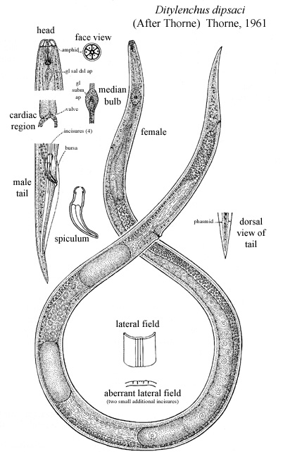
Body marked by transverse striae, about 1 u apart, which are
easily visible under the oil immersion at any point on the body.
Lateral field marked by four incisures. Deirids usually visible near
base of neck. Hemizonid adjacent to excretory pore, about six annules
wide. Phasmids rarely visible and then only from a dorsal or ventral
view on favorable specimens. Amphid apertures on apices of lateral
lips, where they appear as minute refractive dots which can be seen only
from a face view. Spear with strongly developed knobs from which
protrudor muscles lead to the well-sclerotized cephalic framework.
Basal esophageal bulb with the usual three prominent and two inconspicuous,
gland nuclei. Intestine connected to esophageal lumen by a very small
valvular apparatus.
Ovary outstretched, sometimes reaching to
median esophageal bulb, but more often near basal bulb, rarely with one
or two flexures. Oocytes lie largely in tandem and develop into eggs
which are two to three times as long as the body diameter. Rudimentary
posterior uterine branch present, extending about half-way back to anus.
Vulva-anus distance equal to 1 3/4 to 2 1/4 times tail length. Teminus
always acute.
Testis outstretched, with spermatocytes arranged
in single file except for a short region of multiplication. From
a perfectly lateral view the spicula exhibit a sclerotized pattern
that apparently is characteristic of the species, but the proper angle
of observation is so difficult to obtain that the pattern is rarely of
taxonomic value. Bursa rising opposite proximal ends of spicula and
extending about three-fourths the length of the tail. Lateral incisures
ending in a pattern as
illustrated.
Type host: Dipsacus fullonum L., fuller's teasel
Description: Body straight or almost so when relaxed. Lateral field with four incisures. Head unstriated, continuous with adjacent body part. Stylet cone about half of stylet length, knobs rounded. Median esophageal bulb muscular, with thickenings of lumen walls about 4-5 um long. Basal bulb offset or overlapping intestine for a few micrometers. Excretory pore opposite posterior part of isthmus or glandular bulb. Postvulval part of uterine sac about half of vulva-anus distance long or slightly more. Male cloacal alae envelop about three-quarters of tail length. Spicules 23-28 um long. Tail of both sexes conical, always pointed.
Distribution and Economic Importance
Ditylenchus dipsaci is one of the most devastating
plant parasitic nematodes on a wide range of crops. In heavy infestation
crop losses of 60-80% are not unusual; e.g., in Italy up to 60% of onion
seedlings died before reaching the transplanting stage and for garlic crop
losses of about 50% were recorded from Italy and more than 90% from France
and Poland. In Morocco D. dipsaci was found in 79% of seed stocks
of Vicia faba examined (Schreiber, 1977).
(Description- Manual of Agricultural Nematology, Nickle,ed.
1991)
|
|
|